Zoltán Kristóf (ELTE) -from a biologist’s point of view
The European Microscopy Congress was held in Copenhagen from 25 to 30 August. The HSM members were represented by its 8 members only besides Technoorg Linda company (András Szigethy and his team). Five of us had a poster presentation but none of us a talk this time. The Bella Center was the venue for the congress, a huge exhibition complex on the city outskirts. Besides our conference, in the other part of the building, with another entrance, another meeting with a couple of hundred attendants was held during the same period.
Both the venue and the organization were good, however, the app/software that should have provided the information background for the conference was often clunky and its use was not entirely logical. Lectures in the five lecture rooms and a huge plenary hall ran smoothly, and the audience leaving the talks entered the break-out areas including the booths of the companies, posters, several tables for the buffets, and the break-out areas. There was always fruit, seeds, coffee, tea, and water. Nothing was crowded, at least not the places I was in.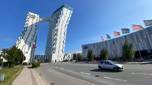
There were sessions on materials science, life science, and imaging, always starting with a plenary in the morning, and the lunch break around noon could be at least partially filled with presentations by one of the exhibiting companies. Unfortunately, it is becoming increasingly rare for exhibitors to provide a lunch package with their presentations. Of course, the life sciences and imaging sessions were the most interesting for me. There was only one presentation on plants, and that was not outstanding. I sat quite diligently through the conference, which for me was all about FIB-SEM, Plasma FIB-SEM, Cryo-CT, vEM, CLEM, Array Tomography, X-ray MicroCT, CLSM, STORM, AI, ML, segmentation.
Anyway, I felt completely rejuvenated, looking around like I did when I first came out west for a conference during the so-called socialist era. Only the excited hope of that time was now replaced by a kind of stoic calm, which was not just because of my age.
There were excellent presentations, demonstrating a lot of work, cooperation, and equipment behind them. The vast majority of PhD students also delivered pretty good presentations. In the technics, there were not many new groundbreaking things. The most interesting for me was the so-called “new” technology. It was amazing to see the structure of the centriole in incredible detail mapped by Ultrastructure Expansion Microscopy. All the beautiful 3D reconstructions I saw were worth it, even if I wasn’t always aware of the technical, and biological significance. But as these techniques become more sophisticated, we are learning more and more about the relationships and dynamics of structures (be they cells, cell organelles, or molecules). As the number of structures in the 3D space under study increases, their complexity becomes more and more difficult to “segment” and follow the individual formulas. Many efforts have been made to develop automatic software to solve this problem, however, even Machine Learning does not seem good enough. I visited a session specifically on this subject, but when I couldn’t understand anything from the fourth lecture either, I gave it up and left. Graphs and complicated mathematical formulas and models are not my thing, but they must be important factors in moving on. Artificial intelligence has been used to generate fluorescent images of tissue and staining and they were indistinguishable from the real thing. Of course, this required the creation of a huge database (www.proteinatlas.org).
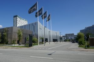
Still, there was time to look around the city in the evenings, and Copenhagen – especially for those who like modern architecture – is a special experience.
Extra points, added by a material scientist, Zsolt Czigány (HUN-REN EK-MFA)
Dániel Olasz and Márk Windisch PhD students from ELTE PhD School, and myself represented the Hungarian material scientists. All of us had poster presentations. I had a good time at the conference. Quite a lot of people (about 8) showed up at my poster. The vendors had a wide variety of products on display. I liked the presentation of Thermo Fisher’s new “Iliad” microscope and visited another microscopic demo, too.
 I have a mixed feeling about the presentations because it was common for invited or plenary speakers to present as if they were talking only to those who know about the same things they do, however, the situation was far from that.
Luckily, there was a plenary lecturer who thought of everybody, too. So I understood the Friday plenary lecture on the connectivity of neural networks better than the previous day’s lecture, which was a more technical topic.
Ádám Soós, PhD student (SE Anatomy), winner of the MMT 2024 Young Lecturer Award. His participation was supported by HSM, too.
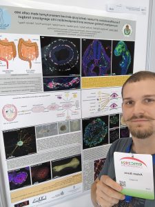 The event was of outstanding scientific and professional quality and provided many opportunities to learn about new technologies and methods in microscopy. I presented my results on the second day of the event. The topic (Transdifferentiation of human dental pulp-derived mesenchymal stem cells into neurosphere forming neurons and transplantation into aganglionic hindgut) was discussed with many interested colleagues and I received valuable feedback. This not only can contribute to the development of my presentation but also to the development of further research directions.
During the conference, I also met several industry players, which may contribute to my learning new techniques. I also attended two workshops that could be particularly useful in the future. The Oxford Instruments workshop was about electron microscope detectors, while the TESCAN workshop was on the company’s latest electron microscope and its applications. I also participated in the demonstration of two new software. The Arivis (Zeiss) software offers new possibilities in image processing, particularly in handling large amounts of data and three-dimensional imaging, the Amiro (ThermoFisher Scientific) software provides novel solutions in data analysis and microscopic imaging.
The 2024 European Microscopy Congress provided an incomparable possibility for me scientifically, professionally, and socially. The new knowledge, contacts, and technological developments will all contribute to furthering my future research.